IN THIS ISSUE...
Welcome to the first edition of our quarterly newsletter!
2017 has arrived and we are excited to begin this edition with the New Year. Our goal is to provide valuable and relative content regarding Mobile Imaging Services, utilizing the information to reach your own goals to grow and succeed.
You’ll find that it is filled with educational information, helpful tips, regulatory updates and articles on MRI safety. We want this newsletter to be valuable for you, so please share your feedback and suggestions for future editions.

Bright Idea of the Month: Why Mobile MRI? Patient Cards

Handing out these information cards to our patients was a great educational tool and answered their questions proactively
A mobile MRI is, technologically, the same as a fixed MRI. Mobile MRI allows our facilities to offer services right in your community that normally could not be offered. It has allowed us to meet rising patient demand while avoiding delays due to construction and renovation. There is dramatically shorter installation time with fewer logistics issues.
Mobile MRI provides:
- A complete and flexible solution
- Superb patient comfort and efficient workflow
- A way to expand services offered
- Less travel for patients
- Flexible fixed schedules and/or on-demand service
- Ability to share services between multiple sites
- Capability to change locations based on patient volume
In adherence to national patient quality and safety standards, every mobile MRI we use is ACR (American College of Radiology) accredited and operated by technologists registered by the ARRT (American Registry of Radiologic Technologists).
Our staff will be glad to answer any questions you may have.
Thank you for choosing us; it is our pleasure to be a part of your healthcare team.
Accreditation of Advanced Diagnostic Imaging Providers
Providers of Advanced Diagnostic Imaging (ADI) in the outpatient setting are required to receive accreditation through a CMS approved agency in order to receive Medicare reimbursement. This requirement was set forth by the Medicare Improvements for Patients and Providers Act of 2008 (MIPPA), and went into effect January 1st 2012. ADI by definition of MIPPA is "advanced diagnostic imaging procedures such as diagnostic magnetic resonance imaging (MRI), computed tomography (CT), and nuclear medicine imaging procedures, such as positron emission tomography (PET). ADI procedures do not include x-ray, ultrasound, fluoroscopy procedures or diagnostic and screening mammography."
Many private payers have followed suit and now have certain requirements for reimbursement of ADI. Most default to the CMS approved agencies for accreditation, but it is always a good idea to consult with your payers on their specific requirements.
Currently, CMS has approved four ADI Accrediting Organizations. Listed below are the names, contact information and web links for these organizations.
American College of Radiology (ACR)
1891 Preston White Drive
Reston, VA 20191-4326
Local telephone number: 703-648-8900
Toll-free telephone number: 800-770-0145
Email address: [email protected]
Website: http://www.acr.org/Quality-Safety/Accreditation
Intersocietal Accreditation Commission (IAC)
6021 University Boulevard, Suite 500
Ellicott City, MD 21043
Toll-free telephone number: 800-838-2110
Email address: [email protected]
Website: http://www.intersocietal.org/
RadSite
326 First Street, Suite 28
Annapolis, Maryland 21403
Telephone number: (443) 440-6007
Email address: [email protected]
Website: http://www.radsitequality.com/
The Joint Commission (TJC)
Corporate Office:
Ambulatory Care Accreditation Program
One Renaissance Boulevard
Oakbrook Terrace, IL 60181
Customer Service: 630-792-5800
Ambulatory Care Accreditation Representative: 630-792-5286
Fax Number: 630-792-5005
Email address: [email protected]
Website: http://www.jointcommission.org/
If you need further information, please reach out to your MedQuest account representatives.
Reference:
Tips on Improving and Optimizing Patient Workflow
We are often asked for tips on how to help a site become more efficient and increase their throughput due to the limited time the mobile MRI is on site. Here are three “best-practice” workflow tips that I have observed in the field.
- Prepare your patients: The most productive sites have a standard message that all scheduled patients receive regarding arrival time, dress and expectations. Work with your MRI staff to determine a realistic arrival time to encompass time for check-in, screening and history. Communicate to patients how they should dress for their exam. A lot of time can be saved by not having to remove multiple pieces of jewelry, clothing with metal or clothing that is made of a material that is not safe to be in the MRI scanner.
- Review your schedule: Many sites employ a practice called “scrubbing” the schedule. This review might look for discrepancies between the physician order and scheduled exam for example. Sites might also review answers to pre-screening questions looking for affirmative responses that might preclude examination. Once a question has been identified, the radiologist and/or MRI staff should be consulted for direction. Another activity that can take place during scrubbing of the schedule consists of confirmation calls. This allows the site to confirm appointments and answer questions in advance. Often by doing these things, sites are able to replace exams that would have been canceled on the schedule with a confirmed exam.
- Practice Critical Analysis: Successful sites practice real-time self-critique of their workflow in order to identify obstacles and brainstorm solutions. How often do you as a practice manager find yourself dealing with the same situation week after week? Maybe you lost some patients on the schedule due to not having enough information about an implanted device. Maybe you constantly run behind schedule. Taking the time to seriously reflect on the root causes of these events can lead to improved processes and superior results. Some tips for practicing critical analysis include:
- Try to make time to review events as close to their occurrence as possible. This makes details of the event clearer in everyone’s mind.
- Include input from the entire team (front line, schedulers, MRI staff, Radiologist and Physicians). This approach insures you get a wider focus and inclusion of ideas. It also helps build consensus for change because everyone feels they were a part of the process.
- After implementation, evaluate the effect of changes on the process to confirm success or the need for additional modification.
By putting into place some of these practices you might discover benefits in patient satisfaction and throughput, improved provider relationships and staff satisfaction.
MRI Safety Corner
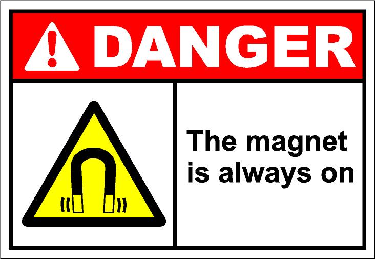
The MRI scanner is a large magnet that is 30,000 times stronger than the earth’s magnetic field. The force increases as a person or a ferromagnetic object moves closer to the magnet. The strong magnetic field of the MR scanner is Always on even when the MR scanner is not in use.
There are many associated risks in the MR environment and reported adverse incidents involving patients, equipment, and personnel. The American College of Radiology (ACR) published in 2002 the initial ACR MR Safe Practices Guidelines. In 2013 the ACR modified and updated numerous standards involving MR Safety. The ACR Guidance Document on MR Safe Practice 2013 was introduced. Tri/Co Mobiles strives to meet the “Gold Standard” in best practices in MR safety and patient care. This month we will be focusing on two important MR safety related topics recommended by the ACR Guidance Document on MR Safe Practices: 2013
There are many associated risks in the MR environment and reported adverse incidents involving patients, equipment, and personnel. The American College of Radiology (ACR) published in 2002 the initial ACR MR Safe Practices Guidelines. In 2013 the ACR modified and updated numerous standards involving MR Safety. The ACR Guidance Document on MR Safe Practice 2013 was introduced. Tri/Co Mobiles strives to meet the “Gold Standard” in best practices in MR safety and patient care. This month we will be focusing on two important MR safety related topics recommended by the ACR Guidance Document on MR Safe Practices: 2013
Safety Issues of the Static Magnetic Field ( ex: 1.5 Tesla or 3 Tesla) Zones
- Zone I: Reception Area/waiting room
- Zone II: Screening interview
- Zone III: Control area with restricted access- On Mobile MR this area is at the entry into the cabin and ALL non-MR personnel must have screening completed and signed by Level II MR-personnel (MR technologist)
- Zone IV: MR scanner room
Patient safety screening form/patient interview
All patients will need to complete a screening form with questions concerning all medical history in addition to any health risk or interferes with the MRI exam. The patient needs to list all surgeries and procedures, including recent colonoscopy and endoscopy. Patients with recent colonoscopy procedures that involve a biopsy may have clips placed in the colon that require a delay of the MR examination.
Examples of items that may create a potential MR health risk to the patient:
- Pacemaker
- Implantable cardioverter defibrillator
- Neurostimulation system
- Aneurysm clip
- Metallic implant
- Implanted drug infusion pump
- Foreign metal objects in eye
- Shrapnel or bullets
- Permanent cosmetics or tattoos (ex: eyebrows)
- Dentures/teeth with magnetic attachments
- Other implants that involve magnets (ex: cochlear implant)
- Medication patch (ex: Fentanyl patch, Nitroglycerin)
Direct verbal and written communication between MRI staff are critical to maintaining patient care and safety. To prevent any inconvenience to the patient all the above devices or implants should be brought to the attention of the MRI technologist or MR Safety radiologist prior to the patients’ scheduled appointment.
Patient care and safety are at the core of all MRI procedures. In accordance, with the “ACR Guidance Document on MR Safe Practices: 2013” all MRI staff must take responsibility for providing a MRI-safe environment for staff and patients. Attention to detail is significant to maintaining MRI safety and patient care.
Everyone must know: